Hopefully after reading this chapter overview, you’ll feel inspired to get both those physical and MCAT gains! Though we suggest focusing on the latter for the time being, these understandings and insights may even help you when you’re making progress at the gym as you’ll really understand the science behind your strength gains!
Working in synergy with the nervous system, it’s amazing to see all the musculoskeletal system can allow for our body’s movement from simple tasks such as walking to more complex one’s like a football player making a one handed catch.
This chapter overview will introduce the basics of the musculoskeletal system, including the main cells, divisions, and so on. Let’s get started!
Musculoskeletal System on the MCAT: What You Need to Know
Topics on the musculoskeletal system will be tested on the biology section of the MCAT and can appear both as passage based and fundamental discrete questions.
Similar to other topics, it’s difficult to narrow down how many musculoskeletal system questions will appear on the MCAT. However, on average, try to expect around 2-4 questions.
Introductory biology account for 65% of the content covered in the Biological and Biochemical Foundations of Living Systems section (Bio/Biochem) and 5% of content tested in the Chemical and Physical Foundations of Biological Systems (Chem/Phys), and 5% of material on the Psychological, Social, and Biological Foundations of Behavior (Psych/Soc).
Important Sub-Topics: Musculoskeletal System
A big component when understanding the musculoskeletal system is its interplay with other organs systems such as the nervous and endocrine system.
Oftentimes, these outside systems give the stimuli to coordinate all the functions and physiological control of the musculoskeletal system.
1. Functions of the Musculoskeletal System
The main roles of the musculoskeletal system include the coordination of the body’s movement and providing support and protection for the body’s internal tissues and organs. The muscles and bones work synergistically to coordinate movement, as muscles will attach to bones and contract, causing the bones and the body to move.
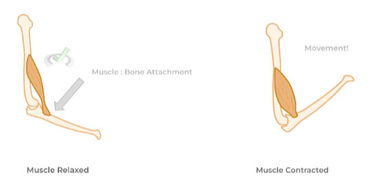
(Coming Soon!) Full Study Notes : Functions of the Musculoskeletal System
For more in-depth content review on the musculoskeletal system, check out these detailed lesson notes created by top MCAT scorers.
2. Types of Muscle: Skeletal, Cardiac, & Smooth
Before delving into the types of muscle, recall that muscles are organs composed of many different types of tissues and cells, with muscle tissue being the obvious main component.
With this, there are 3 main types of muscle tissue (and these muscles) distributed through the body: skeletal, cardiac, and smooth muscle.
Though they all perform muscle contract, they differ in a couple of factors including nuclei number, striation patterns, type of nervous system regulation, etc. Below is a table which summarizes the main differences between the 3 types of muscle.
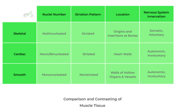
You’ll see that these specific characteristics attributed to each muscle type serves a purpose. For example, the multinucleation of skeletal muscle is due to energy demands required by skeletal muscles for day to day functions!
(Coming Soon!) Full Study Notes : Types of Muscles
For more in-depth content review on the different types of muscle tissues, check out these detailed lesson notes created by top MCAT scorers.
3. Hierarchical Structural of Muscle
Another important concept to keep in mind when studying muscles is that there is a hierarchical organization of the components that make up the muscle. Let’s use an analogy to give a better picture!
The hierarchical muscle organization is similar to how the NBA is organized: from big to small. For example the NBA is divided into 2 conferences. Within each conference are 16 teams. Going even further, each team is divided into +20 individual players and coaches.
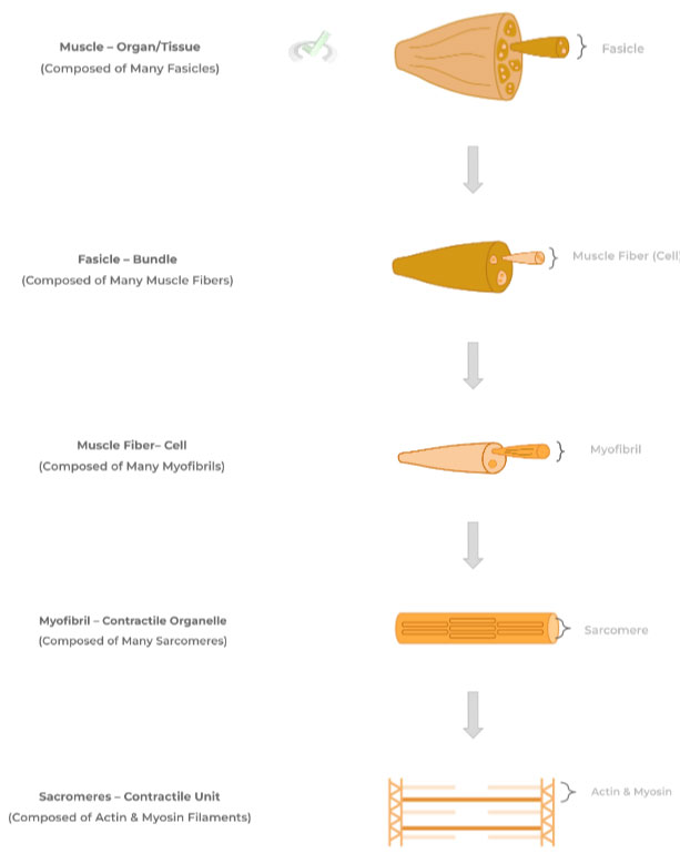
Hopefully the diagram above gives you a better idea and illustration of the hierarchical organization concept!
(Coming Soon!) Full Study Notes : Hierarchical Structure of Muscle
For more in-depth content review on the hierarchical organization concept, check out these detailed lesson notes created by top MCAT scorers.
4. Muscular Contraction: Processes
To initiate muscle contraction, muscle fibers are innervated by somatic neurons delivering motor signals! In order to relay this information, somatic neurons and muscle fibers form a special synapse called the neuromuscular junction. It’s essentially identical to a neuronal synapse, except that the muscle fiber replaces the postsynaptic neuron dendrites.
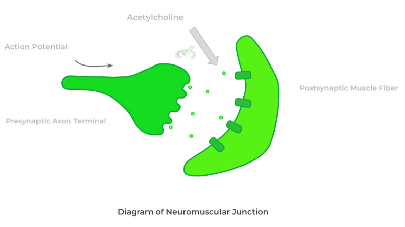
During innervation, an excitatory action potential travels down the axons until it reaches the presynaptic axon terminal. This results in the release of the neurotransmitter acetylcholine which diffuses across the synapse to bind to receptors on the muscle fiber.
Binding results in the depolarization of the muscle fiber, triggering more intracellular processes eventually leading to contraction.
In a more detailed sense, contraction results from the shortening of the sarcomeres, the main contractile unit of the muscle fiber, which are composed of thin, actin and thick, myosin protein filaments.
The sliding filament theory helps to explain how sarcomeres shorten to cause contraction: upon innervation, the myosin filaments head interact and bind with the actin filaments. The myosin heads then “pull” the actin filaments towards the center, shortening the sarcomere and causing contraction.
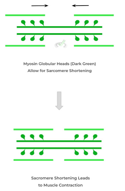
Though they are more specifics, this is the basic overview and idea behind sarcomere shortening and muscle contraction!
(Coming Soon!) Full Study Notes : Muscular Contraction: Processes
For more in-depth content review on the processes involved in muscle contraction, check out these detailed lesson notes created by top MCAT scorers.
5. Anatomy of the Skeletal System and Bone Structure
The skeletal system has 2 main subdivisions: the axial and the appendicular skeleton.
The axial skeleton is primarily composed of the skull, thoracic cage, and vertebral column.The appendicular skeleton is composed of the bones that extend from the axial skeleton such as the bones of the upper arm, lower leg, etc.
One way that might help you conceptualize this is that the axial skeleton comprises the bones that protect the central nervous system!
Though there are many different bone types in the skeletal system, you’ll most likely be tested on the anatomy of the appendicular, long bones such as the femur. Long bones have 2 main divisions: the diaphysis, which is the bone’s long shaft and the epiphysis, which is the bone’s ends.
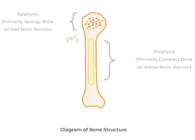
As shown above, there are two types of bone tissue: compact and spongy bone. Both are composed of osteocytes (primary bone cell) and bone matrix, but differ in their arrangements.
Compact bones are composed of osteons, where osteocytes and bone matrix are arranged in concentric circles which form a cylindrical tube. These tubes have various channels where blood vessels enter to supply nutrients and expel waste. Osteons are grouped tightly together, giving the bones their structural rigidity.
Contrarily, spongy bones are not composed of osteons but rather have their osteocytes and bone matrix arranged in what are called trabeculae, which have a more lattice-like arrangement. As shown, the trabeculae of spongy bone contain much more crevices, where the red bone marrow and blood vessels are situated.
Recall that bone is a type of connective tissue, composed of a hydroxyapatite matrix (which functions as the ground substance) and collagen proteins. In addition to osteocytes, osteoblasts and osteoclasts are also embedded within the bone matrix.
The hydroxyapatite matrix is composed of many different types of inorganic molecules, most notably calcium and phosphate! It’s the role of the osteoblasts and osteoclasts to remodel the bone matrix.
Osteoblasts will aid in bone formation, also known as bone deposition, by secreting the collagen and inorganic molecules needed for the matrix. Osteoclasts oppose this function by causing bone degradation, also known as bone resorption.
(Coming Soon!) Full Study Notes : Anatomy of the Skeletal System and Bone Structure
For more in-depth content review on skeletal system anatomy and bone structure, check out these detailed lesson notes created by top MCAT scorers.
6. Endocrine Control of the Skeletal System
In addition to its role in providing bodily support and movement, the bones of the skeletal system are involved in the homeostasis of blood calcium levels! This is due to their role in calcium storage within the hydroxyapatite bone matrix!
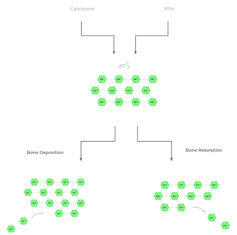
Recall that the main hormones involved in blood calcium homeostasis are calcitonin and the parathyroid hormone (PTH), which work to decrease and increase blood calcium levels respectively.
This makes sense when understanding what each hormone promotes: calcitonin will promote bone deposition as calcium will be uptaken from the bloodstream for the bone to store within the hydroxyapatite matrix. Conversely, PTH promotes bone resorption and utilizes the released calcium ions to increase blood calcium levels.
(Coming Soon!) Full Study Notes : Endocrine Control of Skeletal System
For more in-depth content review on endocrine function within the skeletal system, check out these detailed lesson notes created by top MCAT scorers.
Important Definitions and Key Terms
Below are some high yield definitions and key terms to refer to when reviewing the musculoskeletal system!
Term | Definition |
|---|---|
Fascicle | Bundles of muscle fibers; many fascicles will come together to form muscle |
Myofibril | Main contractile organ found within muscle fibers |
Sarcomere | Main contractile unit found within myofibrils |
Neuromuscular Junction | A specialized synaptic connection made between the axon of a somatic neuron and a muscle fiber |
Sliding FIlament Theory | Theory which explains muscle contraction via the shortening of sarcomeres through the interaction of the actin and myosin filaments |
Epiphysis | Term to describe the bone’s end, primarily composed of spongy bone and red bone marrow |
Diaphysis | Term to describe the bone’s long shaft, primarily composed of compact bone and yellow bone marrow |
Osteons | Concentric, tubular arrangement of osteocytes and bone matrix within compact bones |
Osteoblast | Bone cell which promotes bone formation/deposition |
Osteoclasts | Bone cell which promotes bone degradation/resorption |
Additional FAQs - Musculoskeletal Systems on the MCAT
A. Do You Need To Know Bones for the MCAT?
B. What are 6 Main Functions of the Musculoskeletal System – MCAT?
Additional Reading Links (Coming Soon!) – Study Notes for Muscoskeletal Systems on the MCAT
Additional Reading: MCAT Biology Topics:
- Cells on the MCAT
- Digestive Systems on the MCAT
- Embryogenesis and Development on the MCAT
- Endocrine Systems on the MCAT
- Excretory Systems on the MCAT
- Genetics and Evolutions on the MCAT
- Immune Systems on the MCAT
- Nervous Systems on the MCAT
- Cardiovascular Systems on the MCAT
- Reproduction on the MCAT
- Respiratory Systems on the MCAT


 To help you achieve your goal MCAT score, we take turns hosting these